

This article is reviewed, corrected, and approved by: Dr. Joshua Collins M.D. | MRCP। FRCP
Mouth larva may sound like something out of a horror movie, but it's an unfortunate reality for some. Fly larvae from the sarcophagidae family (flesh fly) cause oral myiasis frequently occur by invading open sores in the mouth and laying their eggs there. These larvae target the wound area and feed on necrotic tissue while preserving healthy tissue, making them an effective solution for wound care.
In this article, we'll take a closer look at mouth larva or oral myiasis – what it is, what oral myiasis causes and its symptoms, how oral myiasis is treated, and how to prevent it from happening.
What is Mouth Larva?
Gums and extraction wounds (open wounds) are vulnerable to invasion by larval fly species. In some cases, it is possible for oral larvae to infiltrate the body via the mouth or other entry points.
The larvae of flies are commonly referred to as "maggots" or "grubs." They are small, white, and have a worm-like appearance. Once they enter the body, the larvae lay eggs and quickly multiply, also causing infection if left untreated on time. These parasites consume either the living or dead tissue of their hosts in order to survive.
Oral myiasis is most common in people who are malnourished, immunocompromised, or living with poor mouth or dental hygiene conditions. It is also more common in warm climates as the fly larvae need moist environments to survive and reproduce.
If you think you may have an oral myiasis infection, it is essential to seek medical advice and treatment as soon as possible to prevent serious complications. Let's see what causes mouth larvae.
Causes of Mouth Larva or Oral Myiasis
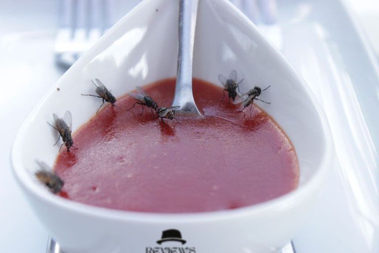
Flies causing myiasis belong to a group of insect dipterans, which includes several families, Calliphoridae and Sarcophagidae. Generally, these flies are attracted to wounds, open sores, or if you have a previous nosocomial infection.
They lay their eggs in these areas, and their larvae hatch and burrow into the skin or tissue. Once inside the body, they feed on the surrounding living tissue, and as they mature, they can cause further damage by migrating throughout the body.
In some cases, oral myiasis can be caused by contaminated food. When an infected fly lays eggs in ingested food, the larvae get stuck in the mouth's oral tissues and feed on mouth and throat mucous membranes. This can cause pain and irritation.
Oral myiasis can also myiasis occur of improper hygiene practices or inadequate medical care. For example, if a wound is left untreated, it can become a breeding ground for flies to lay their eggs if you eat uncooked or unsafe food.
Similarly, plaque buildup can invite flies to lay eggs if a person has poor oral hygiene. As well as, if a person does not receive adequate medical care for a wound, this can lead to further complications that may attract flies.
Types of Mouth Larvae
Mouth larvae are the larvae of the family Chrysomya bezziana and Sarcophagidae, which are flies. And there are many different species responsible for primary oral myiasis. Some of the most common types of mouth larva include:
Cochliomyia hominivorax
This type of larva is commonly referred to as "human botflies" and is known to cause an infestation in both humans and animals. They lay their eggs near wounds and open sores and are most commonly found in tropical and subtropical countries.
Cordylobia anthropophaga
This fly species is commonly known as "mango fly" or "tumbu fly" and is typically found in African regions. The worst case of mangoworms in humans involves the larvae burrowing deep into the skin, causing tissue damage, infections, and even sepsis.
Lucilia sericata
This fly species is often called "green bottle fly" and is typically found in temperate climates. The female lays eggs on wounds and open sores in human and vertebrate animals. The larvae can quickly penetrate the skin and cause an infestation if not treated properly.
Muscina stabulans
This species of fly is known as "stable flies" and is typically found in temperate climates. The female lays her eggs on clothes or bedding, and if left in contact with the skin for a long period of time, the larvae can penetrate into the skin and cause an infestation.
As well as these most common types of mouth larva, other species can also cause oral myiasis. If you suspect you have been infected with any mouth larva, you should seek medical attention immediately.
Risk Factors of Mouth Larvae
Here are some risk factors of maggots in mouth or mouth larva are
- Poor oral hygiene.
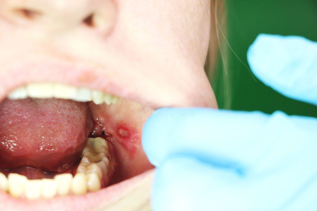
- Open wounds or ulcers in the mouth (mouth ulcer).
- Malnutrition.
- Immobility.
- Poor living conditions.
- Reduced cognitive function.
- Substance abuse.
- Diabetes.
Symptoms of Oral Myiasis

Fly larvae are the culprits behind oral myiasis, a condition characterized by unpleasant side effects. Symptoms of oral myiasis can vary depending on the type of fly larvae involved. Common signs and symptoms include:
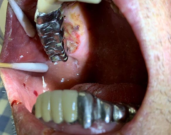
• Oral discomfort: Pain, swelling, and tenderness in the affected area can be present, like gingivitis. For example, cutaneous myiasis is a sign of maggots in gums.
• Presence of larvae: Larvae are often visible to the naked eye and may be seen moving or stuck to the mucosa or soft tissue of the mouth.
• Inflammation: Inflammation and redness around the affected area may also be present.
• Bad breath: Due to the presence of decaying organic material in the affected area, bad odor or breath can be a result.
• Discharge: A yellowish-green discharge may be seen due to the presence of larvae and decaying organic material.
• Difficulty eating and speaking: According to j oral sci, pain and inflammation can make it difficult to eat and speak normally.
Besides, keep in mind that not all cases of oral myiasis will have the same symptoms; myiasis symptoms in humans varies person to person. And some people may experience more severe symptoms than others.
Diagnosis of Oral Myiasis
The most effective way to diagnose oral myiasis is by visual inspection of the affected area. It is important to look for larvae, as well as any other infection-related symptoms may see, such as redness, inflammation, or pus. If the doctor suspects oral myiasis, they may check a swab of the wound and send it for laboratory testing to confirm the presence of the larvae.
If the infection is mild, a doctor may be able to treat the wound simply by removing the larvae. Possible recommendations for antibiotics or antifungal creams can be used under medical supervision. However, antibiotics or antifungal creams may be recommended in some cases to help eliminate the infection and stop it from spreading.
If the condition has caused more severe damage to the dead or living tissues, surgery may be necessary to remove the larvae and repair any tissue damage. In rare cases, the infection can be life-threatening if left untreated.
If you suspect you may have oral myiasis, you may consult an oral and maxillofacial pathology specialist for a proper diagnosis and treatment plan. Based on your requirements, your physician will assess and recommend the optimal plan of action based on individual needs.
How to Treat Mouth Larvae?
Diagnosis for Mild Condition
Many types of treatment are available to treat mouth larvae or oral myiasis. Basically, it depends on the location of the infection and whether it is mild or severe.
In most cases, a medical professional can manually remove the larvae from the affected area. In cases where larvae are deeply embedded, a topical or injectable anesthetic may be used to help with the removal process.
In some cases, anti-parasitic medications may be used to kill the larvae and prevent reinfection. Commonly prescribed medications for this include ivermectin and albendazole. Moreover, antibiotics may be prescribed to treat any secondary bacterial infections that can occur with oral myiasis.
Diagnosis for Severe Condition
In certain situations, sterilized fly larvae can be used to treat persistent, non-healing wounds or sores. This therapy, also known as maggot debridement therapy (MDT), involves the application of sterile maggot infestations area, which can help clean and debride the wound and stimulate tissue growth and growth healing.
No matter what kind of treatment is chosen, maintaining cleanliness and dryness in the afflicted area is essential in avoiding infection recurrence. In order to get rid of any leftover larvae or eggs, it is also required to deep clean all of your clothes and bed sheets in hot water before drying them on a high heat setting.
You Will Be Surprised to Know These Facts About Myiasis
Conclusion
Mouth maggots or oral myiasis is an infestation of the mouth caused by fly larvae, and it can cause painful symptoms such as burning, irritation, and difficulty eating. In tropical areas, you may protect yourself using mosquito nets, window nets, or insect repellant might help you.
You should speak with a doctor if you experience any of these symptoms, as early diagnosis and treatment are essential to successfully treating this condition. While oral myiasis is rare, knowing the signs and symptoms can help you take the necessary steps to treat it.
F.A.Q
Q: What causes mouth larva?
Ans: It may be caused by poor oral hygiene, impaired consciousness, malnourishment, wounds, or lesions in the mouth, like mouth ulcers.
Q: What is míase?
Ans: Miase is caused when fly larvae get into the tissue of a living animal, like a person. It is also known as myiasis.
Q: Is oral myiasis a common condition?
Ans: Incidences of oral myiasis are rare, making it an uncommon medical problem.
Q: How is oral myiasis diagnosed?
Ans: The doctor may order more diagnostic tests to validate the diagnosis and determine how bad the infestation is. Tests such as X-rays, CT scans, laboratory analyses and tests, and blood tests are types of imaging examinations.